Change Language German
Definition
- Optic disc swelling due to increased intracranial pressure
Symptoms
- Neurological symptoms
- Headaches: new, usually severe; worse in the morning; exacerbated by coughing, physical exertion, lying down, or bending forward
- +/- pulsatile tinnitus
- +/- non-specific paresthesias or weaknesses
- +/- other neurological deficits
- Ophthalmological Symptoms
- initially usually normal vision
- Blurred vision / reduced visual acuity
- Hypermetropisation
- Transient visual obscurations
- Photopsia
- +/- diplopia
- Secondary: reduced color and contrast sensitivity, visual field defects (initially typical in the mid-peripheral field)
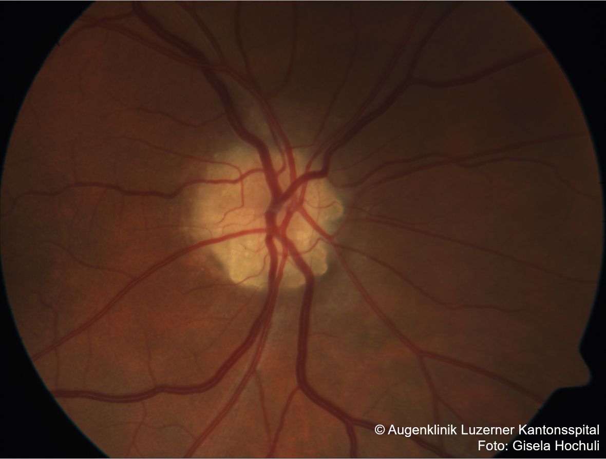
Findings
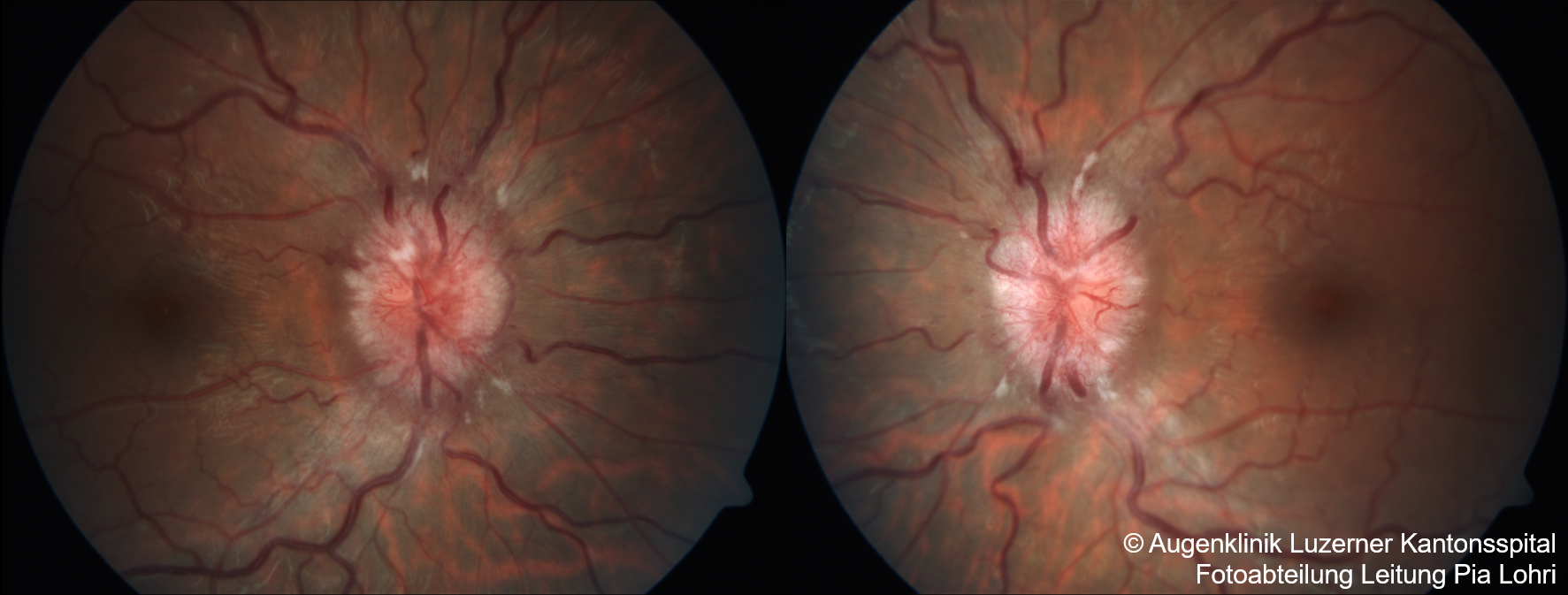
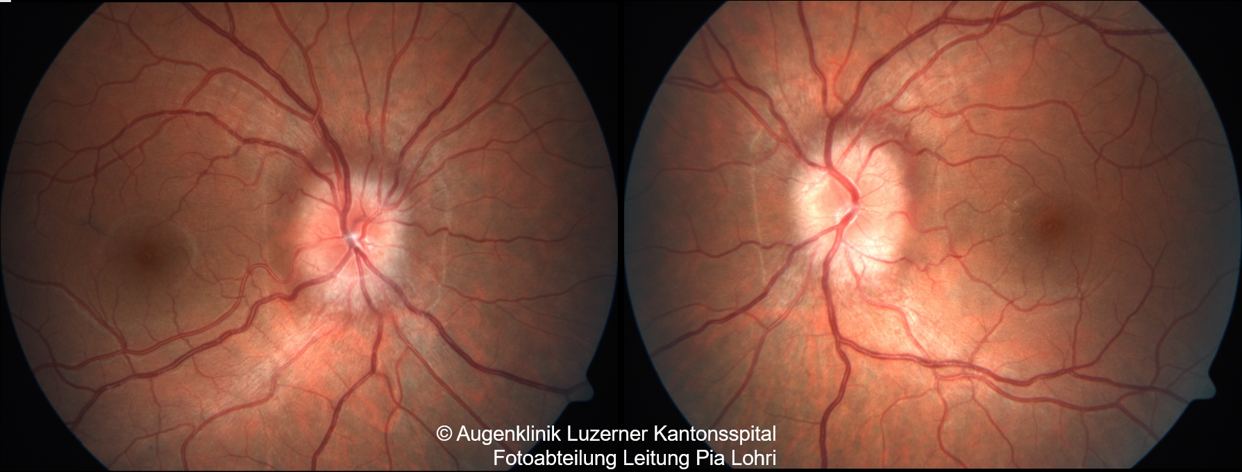
- Papilledema : graded according to Frisén Grading Scale (Grade 1-5)
- Obscuration of vessels around the optic disc, absent optic cup, flame-shaped hemorrhages, spontaneous venous pulse not visible, dilated retinal veins, absent nerve fiber layer reflex
- Usually bilateral, rarely unilateral
- Initially normal afferent visual function (visual acuity, color/contrast vision, no RAPD)
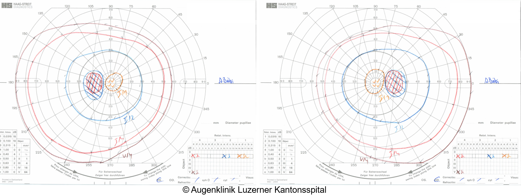
- Visual fields initially usually normal (apart from enlarged blind spots)
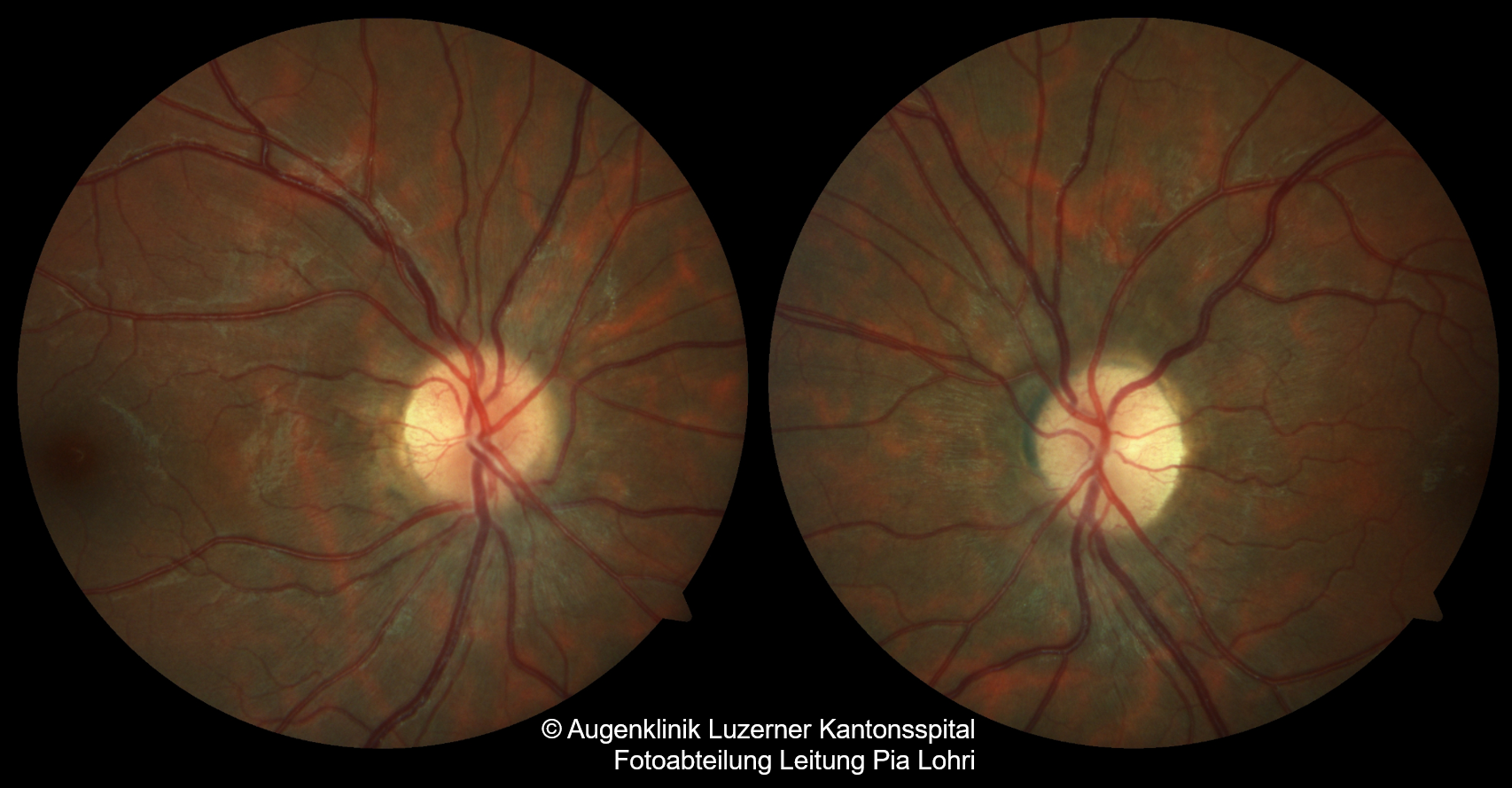
- Secondary optic atrophy after regression of the swelling
Aetiology
- Pseudotumor cerebri = idiopathic intracranial hypertension
- Brain tumors, intracranial hemorrhage, brain abscess
- Brain edema from trauma or metabolic origin
- Small skull in craniosynostosis (very rare)
- Hydrocephalus
- Primary: congenital or early acquired (not in children or adults)
- Secondary communicating or obstructive hydrocephalus (enlarged ventricles visible in imaging)
- Subarachnoid hemorrhage (SAH)
- Meningitis
- Increased cerebrospinal fluid production by choroid plexus tumors (rare)
- Sinus vein thrombosis (often of the superior sagittal sinus and/or transverse sinus)
- Extracranial venous outflow obstruction e.g., obstruction of internal jugular vein or superior vena cava
Differential Diagnoses
- “Pseudo-Papilledema”
- No vascular obscuration or dilation, normal nerve fiber reflex, no haemorrhages, preserved optic cup, usually present spontaneous venous pulse
- E.g., optic disc drusen, ‘crowded disc’/congenital optic disc anomaly, ’tilted disc’
- Optic disc swelling of other etiology, including diabetic papillopathy, anterior ischemic optic neuropathy (AION), papillitis, etc.
Work-up
To distinguish true papilledema from pseudo-papilledema
- Fundoscopy
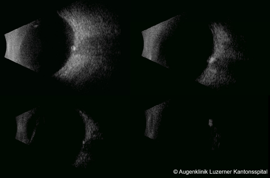
- Ultrasound: Hyperreflectivity of drusen
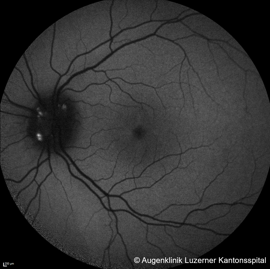
- Autofluorescence: Hyperautofluorescence of drusen
- Fluorescein angiography: No leakage in pseudo-papilledema, only staining
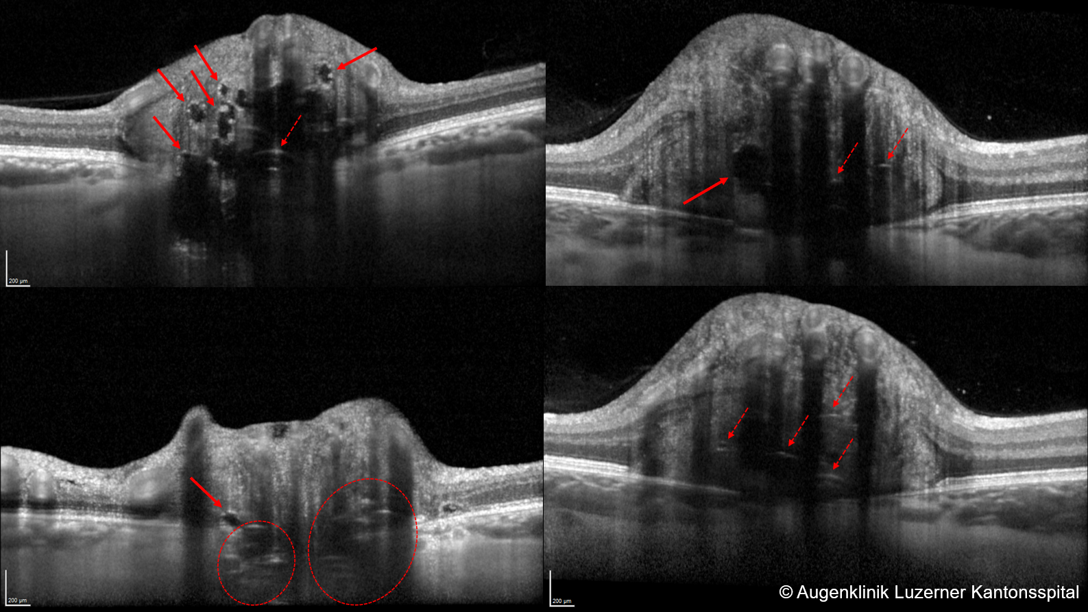
- OCT (EDI images): Possibly visible drusen
- CT: Intracranial space-occupying lesion? Increased intracranial pressure? Hyperreflectivity of drusen?
- MRI: Intracranial space-occupying lesion? Enlarged optic nerve sheaths? Posterior flattening of the globe?
- Lumbar puncture, if above investigations are inconclusive: Increased opening pressure? (normal opening pressure is <25cm CSF in adults or <28cm CSF in children), cerebrospinal fluid analysis
Sources
- EyeWiki Papilledema
- The Wills Eye Manual: Office and Emergency Room Diagnosis and Treatment of Eye Disease; Kalla Gervasio MD, Travis Peck MD et al; Lippincott Williams&Wilkins; 8th Edition (2021)
- Kanski’s Clinical Ophthalmology: A Systematic Approach; John E Salmon MD; Elsevier; 9th Edition (2019)