Change Language German
Aetiology
- Autoimmune process, allergic hypersensitivity reaction Type II
Clinical Presentation
- Age typically 60-80 years, more common in women (female-to-male ratio 2:1)
- Symptoms: insidiously starting or recurrent nonspecific bilateral conjunctivitis, often initially misdiagnosed as dry eyes
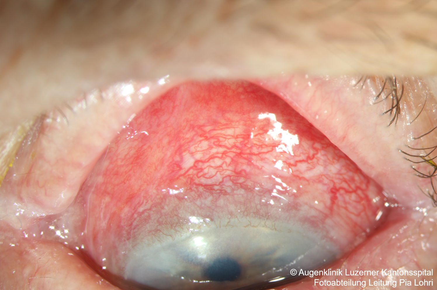
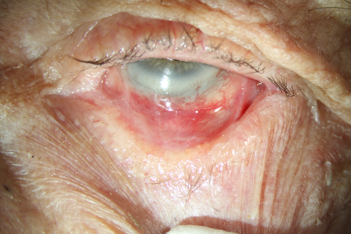
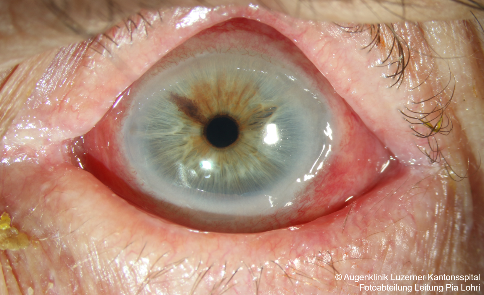
Findings
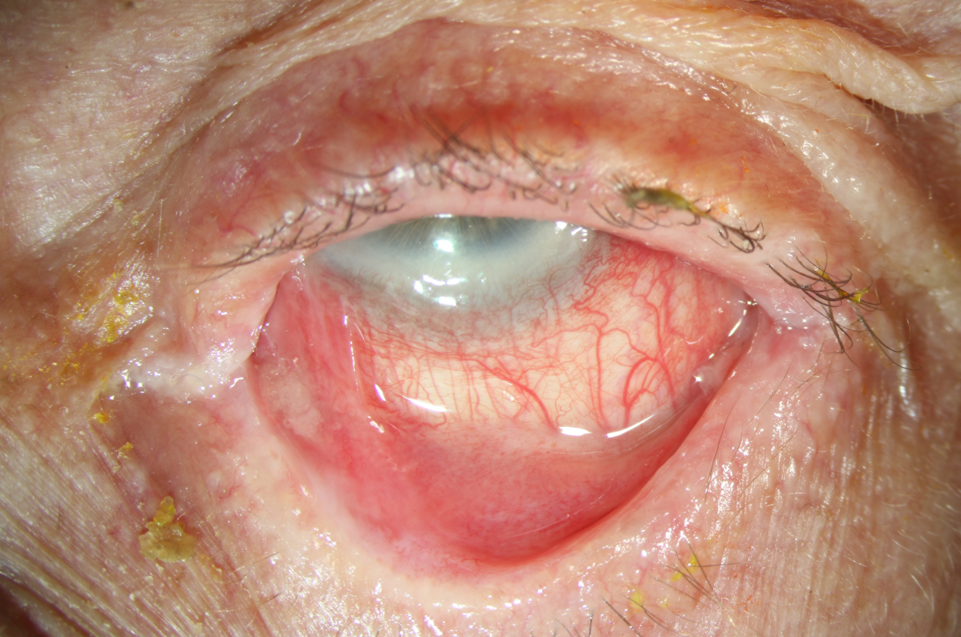
- Conjunctivitis, diffuse hyperemia
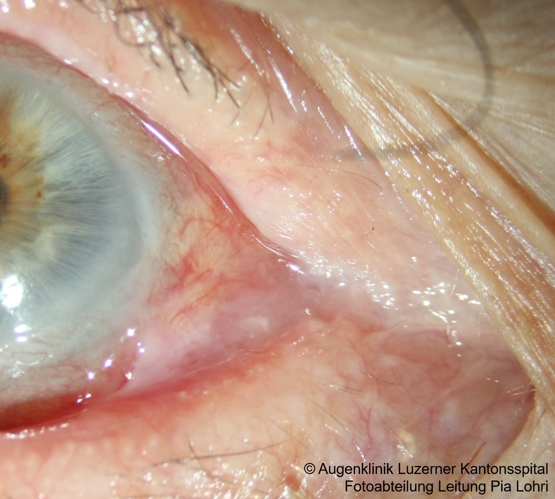
- Subconjunctival fibrosis
- Fornix shortening
- Keratinization of the caruncle/absence of caruncle/plica
- Fibrotic/scarred tear ducts , destruction of goblet cells (-> dry eye)
- Cornea: Epithelial defects, dellen, ulcers, perforation, vascularisation, keratinisation/conjunctivalisation
- Symblepharon (adhesion of the bulbar to the palpebral conjunctiva)
- Ankyloblepharon (adhesion between upper and lower lid)
- Entropium with trichiasis
Foster’s Classification System
- Stage I: Early stage with minimal changes (subepithelial fibrosis, chronic conjunctivitis)
- Stage II: Fornix shortening
- Stage III: Symblepharon
- Stage IV: Ankyloblepharon
Work-up
- Biopsy (histology + immunofluorescence: linear deposits of IgG, IgA, and/or complement C3 along the basement membrane)
Treatment
- Local lubrication (preservative-free), autologous autologous serum eye drops
- Local steroids or topical cyclosporine (Ikervis) against local inflammation (usually not sufficient)
- Systemic therapy/immunosuppressants
- Dapsone
- Methotrexate
- Azathioprine (Imurek)
- Mycophenolate mofetil
- Cyclophosphamide
- For dosages and recommended lab tests, see treatment recommendations from rheuma-net.ch
Differential Diagnoses/ Pseudopemphigoid
- Drug-induced: chronic topical therapy (including Pilocarpine, Epinephrine, Timolol, Idoxuridine)
- No progression after discontinuation of medications, clinical changes especially in the inferior fornix
- Post-inflammatory scarring
- Autoimmune diseases: Sarcoidosis, scleroderma, lichen planus, Stevens-Johnson, dermatitis herpetiformis, epidermolysis bullosa, atopic blepharoconjunctivitis, graft-vs-host-disease
- Trauma: chemical injuries, burns
- Severe blepharoconjunctivitis e.g. in rosacea
- IgA dermatosis (clinically similar presentation!)
Sources
- Valerie P.J. Saw, John K.G. Dart, Ocular Mucous Membrane Pemphigoid: Diagnosis and Management Strategies, The Ocular Surface, Volume 6, Issue 3, 2008, Pages 128-142, ISSN 1542-0124, https://doi.org/10.1016/S1542-0124(12)70281-1
- EyeWiki Ocular cicatricial pemphigoid
- Behandlungsempfehlungen bei Immunsuppression der Schweizerischen Gesellschaft für Rheumatologie
- The Wills Eye Manual: Office and Emergency Room Diagnosis and Treatment of Eye Disease; Nika Bagheri MD, Brynn Wajda MD, et al; Lippincott Williams&Wilkins; 7th Edition (2016)
- Kanski’s Clinical Ophthalmology: A Systematic Approach; Jack J. Kanski MD, Brad Bowling MD; Saunders Ltd.; 8th Edition (2015)