Change Language German
Findings
- Orbit: Enlarged lacrimal glands, involvement of extraocular muscles (double vision, proptosis, restrictive)
- Eyelids: Ptosis
- Conjunctiva: Localised or diffuse yellowish waxy lesions
- Cornea: Dry eye, variant of Lattice corneal dystrophy, Meretoja syndrome
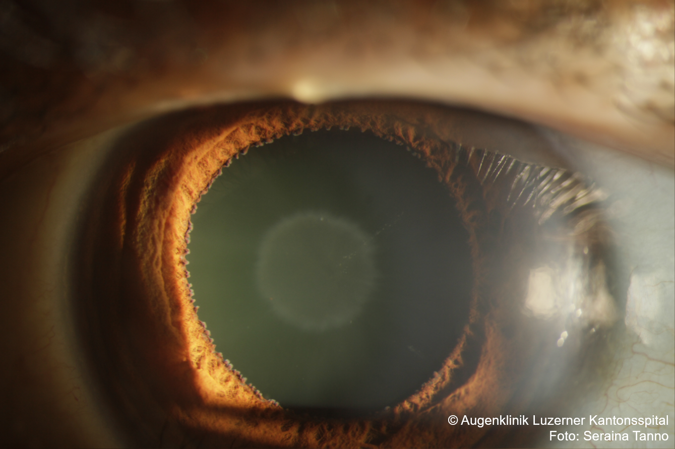
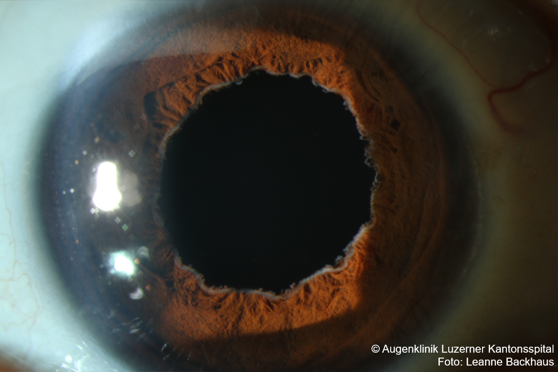
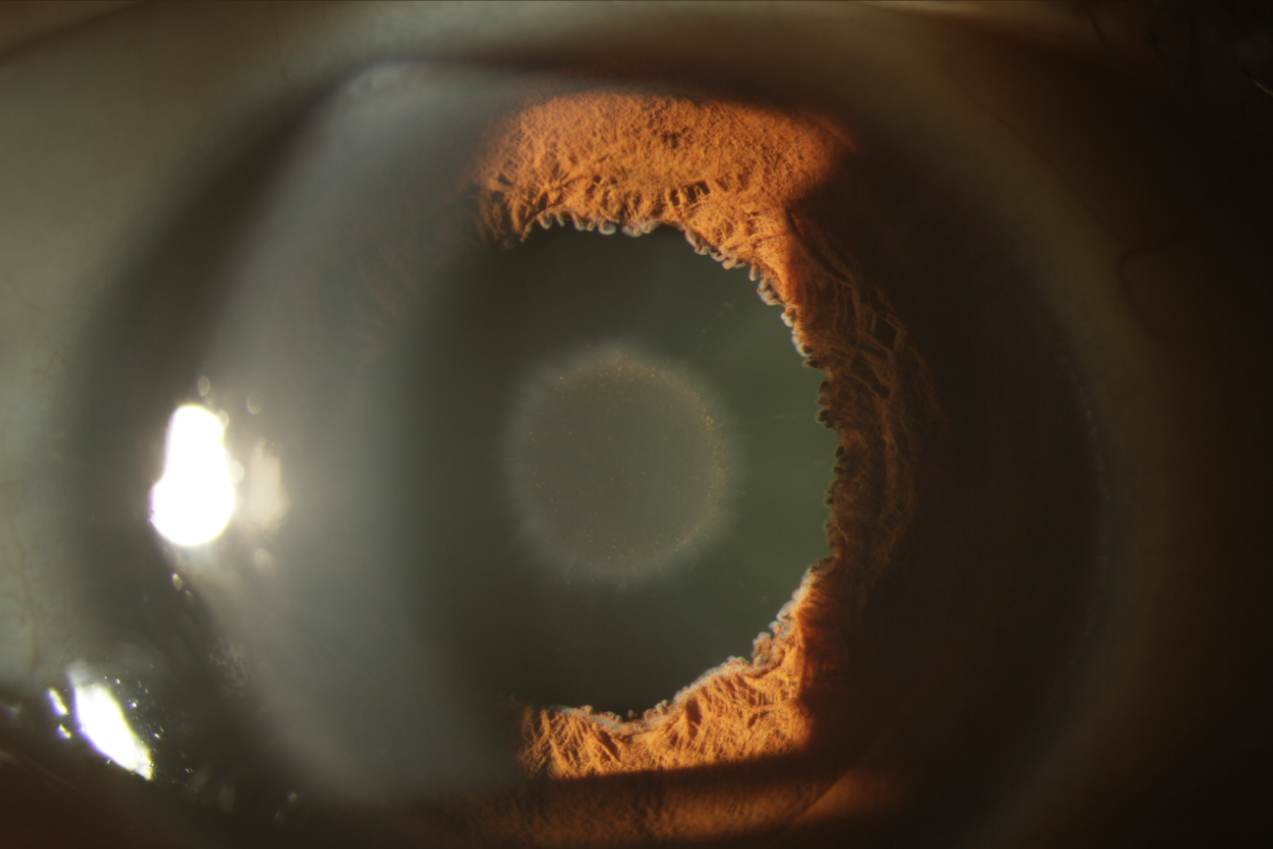
- Iris: Deposits at pupillary margin , fringed/scalloped pupil
- Glaucoma: Secondary due to amyloid deposits in the trabecular meshwork
- Lens: Deposits on the anterior surface of the lens
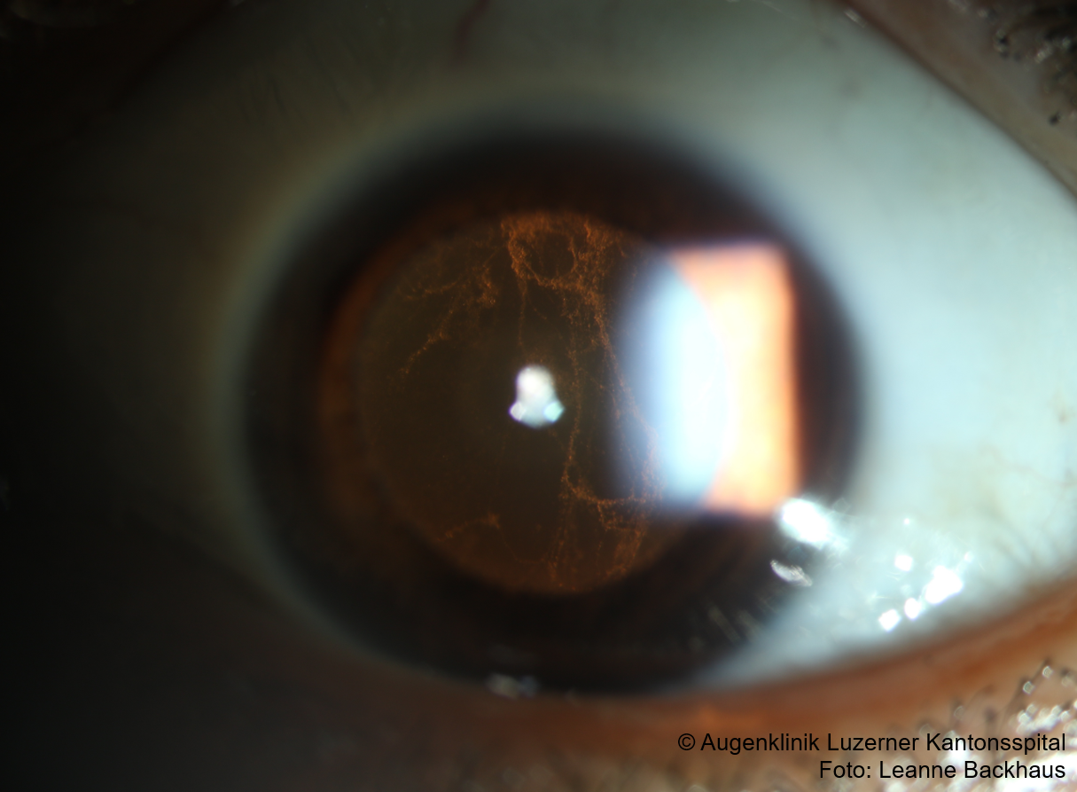
- Vitreous: Glass wool-like opacities (usually visually disturbing)
- Retina: Vitreous tufts adjacent to retinal vessels
- Optic neuropathy (compressive, infiltrative) rare
Work-up
- Family history
- Slit lamp examination including fundoscopy
- Imaging: CT/MRI of the orbit
- Biopsy (conjunctival lesions or lacrimal gland)
- Genetic work-up
Treatment
- Orbit: Consider decompression
- Conjunctival lesions: Lubrication (e.g., Lacrycon AT), +/-excision
- Glaucoma: Topical, surgical
- Lens: Cataract surgery
- Vitreous opacities: Pars plana vitrectomy
- Systemic: Adapted to type of amyloidosis
Sources
- EyeWiki Ocular Amyloidosis
- Kimura A, Ando E, Fukushima M, Koga T, Hirata A, Arimura K, Ando Y, Negi A, Tanihara H. Secondary glaucoma in patients with familial amyloidotic polyneuropathy. Arch Ophthalmol. 2003 Mar;121(3):351-6. doi: 10.1001/archopht.121.3.351. PMID: 12617705.