Change Language German
Aetiology
- Lesion of the oculo-sympathetic pathway
- Causes
- Central (1st neuron): Brainstem tumor or infarct, spinal tumor, Multiple Sclerosis, cervical spondylosis
- Preganglionic (2nd neuron): Pancoast tumor, dissection of the internal carotid artery, iatrogenic (neck or thoracic operations); children: Neuroblastoma, Lymphoma
- Postganglionic (3rd neuron): Dissection of the internal carotid artery, tumor or inflammation of the cavernous sinus, iatrogenic (neck operations), autonomic trigeminal cephalgias (e.g., Cluster headache, paroxysmal hemicrania)
Findings
- Miosis, dilation lag, more pronounced anisocoria in the dark
- Mild upper lid ptosis, inverse ptosis of the lower lid -> Pseudoenophthalmos
- Anhidrosis
- Heterochromia in congenital Horner syndrome
Work-up
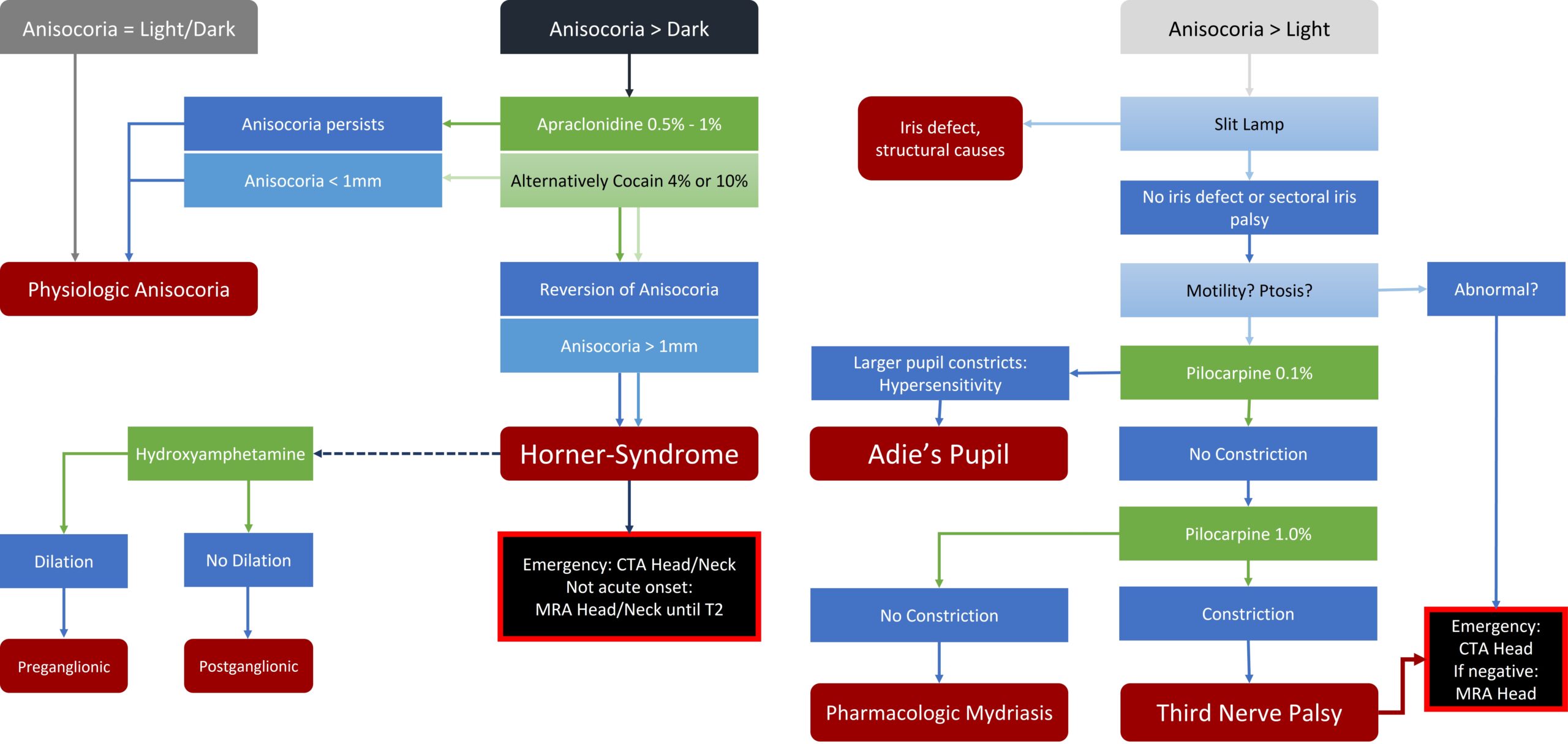
- Iopidine (apraclonidine) 0.5% or 1%, 1x (evaluation after 30-45 minutes)
- Reversal of anisocoria in Horner syndrome
- Contraindicated in children <1-2 years
- Diagnostic gap: Can be falsely negative in very acute cases!
- Cocaine Test: Cocaine 10%, 1x (evaluation after 60 minutes)
- Dilation in a healthy pupil, no effect/no mydriasis in Horner pupil
- Anisocoria <1 mm: Horner syndrome unlikely
- Anisocoria >1 mm: Horner syndrome likely
- mainly in children <2 years (apraclonidine contraindicated!)
- No diagnostic gap
- Dilation in a healthy pupil, no effect/no mydriasis in Horner pupil
- Hydroxyamphetamine 1%, 1x (evaluation after 45 minutes)
- To differentiate post/pre-ganglionic Horner syndrome
- Dilation if lesion is in the 1st or 2nd neuron; no dilation with a lesion in the 3rd neuron
- To differentiate post/pre-ganglionic Horner syndrome
- Phenylephrine 1%, 1x
- To differentiate post/pre-ganglionic Horner syndrome (alternative to Hydroxyamphetamine test)
- Dilation with a lesion in the 3rd neuron; no dilation with a lesion in the 1st or 2nd neuron
Imaging
- In case of acute Horner syndrome:
- immediate CT Angiography (head + neck) to rule out a carotid dissection
- In case of long-standing Horner syndrome:
- MRI Angiography head + neck (up to T2) within 1(-2) weeks
Sources
- EyeWiki Anisocoria
- The Wills Eye Manual: Office and Emergency Room Diagnosis and Treatment of Eye Disease; Nika Bagheri MD, Brynn Wajda MD, et al; Lippincott Williams&Wilkins; 7th Edition (2016)
- Kanski’s Clinical Ophthalmology: A Systematic Approach; Jack J. Kanski MD, Brad Bowling MD; Saunders Ltd.; 8th Edition (2015)