Change Language German
Introduction
- 3 main types of electroretinograms (ERGs)
- Full-field ERG (ffERG): Illuminates the entire retina uniformly
- Pattern ERG (PERG): Utilizes a contrast stimulus, typically an alternating checkerboard pattern
- Multifocal ERG (mfERG): Provides a topographic map of cone system function over approximately 50° of the retina
- Characteristics of Full-field ERG
- DA = Dark Adaptation
- LA = Light Adaptation
- 0.01 / 3.0 / 10.0 = Flash intensities, indicating the strength of the light flash
- a-wave: Response of photoreceptors (negative wave)
- b-wave: Response of Müller cells and bipolar cells (positive wave)
- c-wave: Response of the retinal pigment epithelium (RPE), 2-4 seconds delayed, (positive wave)
- occurs only in dark adaptation
- Implicit time: Time from light stimulus to the peak of the b-wave (in milliseconds)
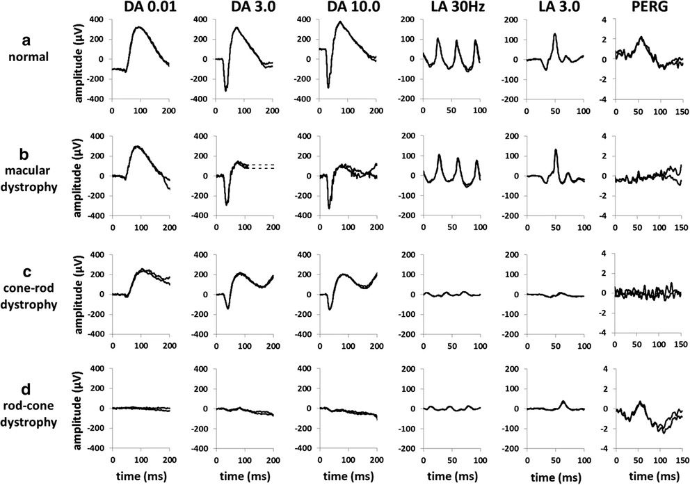
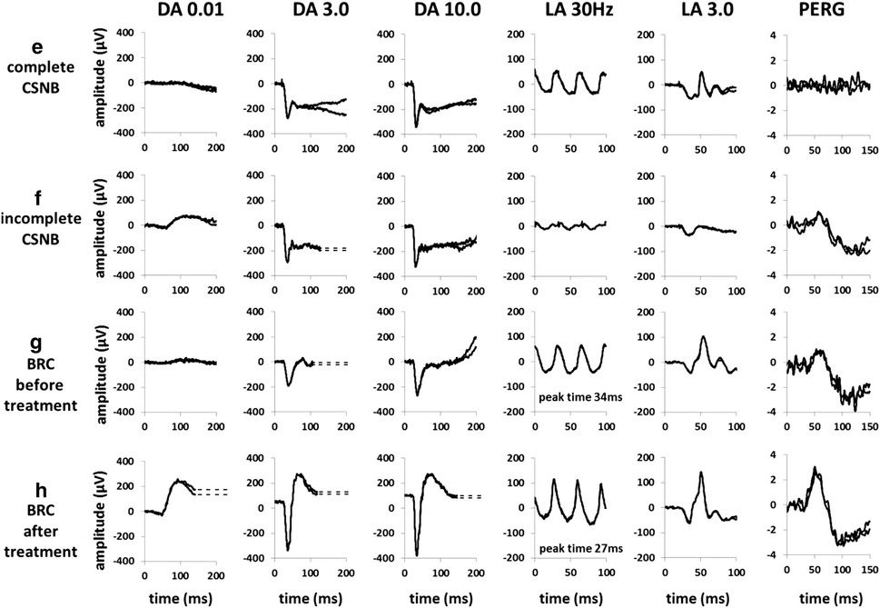
Overview ERG und PERG¹
- DA 0.01: Rod-specific; b-wave, cannot differentiate between photoreceptors and inner retinal layers.
- DA 3.0: Mixed rod-cone response; includes both a- and b-waves.
- DA 10.0: a-Wave indicates photoreceptor function; differentiates between photoreceptor dysfunction and inner retinal dysfunction.
- If DA 0.01 is reduced:
- DA 10.0 a-wave reduced? -> photoreceptor dysfunction
- DA 10.0 b-wave reduced? -> inner retinal dysfunction
- If DA 0.01 is reduced:
- LA 30 Hz: “photopic flicker” tests cone function
- LA 3.0: “photopic single-flash” a-wave corresponding to cone photoreceptors and off-bipolar cells, b-wave with on/off-bipolar cells
- PERG: Pattern ERG: contrast stimulus, typically an alternating checkerboard pattern at consistent brightness
- Must be focused on the macula
- e.g. LHON
- Main components
- P50: Amplitude allows objective assessment of macular function
- N95: Measures central retinal ganglion cell function (RGCs)
- Additional component: N35
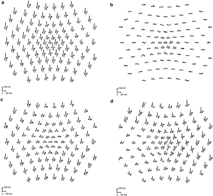
Multifocal ERG¹
- Topographic representation of the cone system over approximately 50°
- Good fixation is important
- e.g. for early detection of hydroxychloroquine retinopathy
- a) Normal finding; b) Retinitis pigmentosa; c) Macular dystrophy; d) Enlarged blind spot in eccentric nasal retinal dysfunction
Indication
- Diagnosis of generalized retinal degenerations
- In cases of suspected reduced visual acuity and presence of nystagmus at birth
- Measuring retinal function in cases of opaque media
- If functional visual loss is suspected
Examples
- Central Retinal Artery Occlusion (CRAO): Normal a-wave (as the photoreceptor layer is supplied by the choroid), missing b-wave
- Ischemic Central Retinal Vein Occlusion (CRVO): Reduced amplitude of b-wave, extended implicit time
- Retinitis Pigmentosa: Reduced amplitude (usually of the b-wave) and extended implicit time; in advanced stages -> no rod and cone response to bright light stimuli
- Multiple Evanescent White Dot Syndrome (MEWDS): Reduced a-wave
- Glaucoma, Congenital Rubella, Optic Atrophy/Neuropathy: Normal ERG (as ganglion cells are affected)
Electrooculogram (EOG)
- Measures corneoretinal potential
- Assesses the function of the RPE and the interaction of photoreceptors with RPE
- Arden Ratio: Maximum potential height in light divided by minimum potential height in darkness
- Normal: > 1.85/ > 185%
- Pathological: < 1.65/ < 165%
- The ERG is pathological in all cases where the EOG is abnormal except:
- Normal ERG, pathological EOG:
- Best’s disease as a classic example
- Pattern dystrophies, Chloroquine retinopathy
- Pathological ERG, normal EOG:
- X-linked retinoschisis, CSNB (congenital stationary night blindness)
- Normal ERG, pathological EOG:
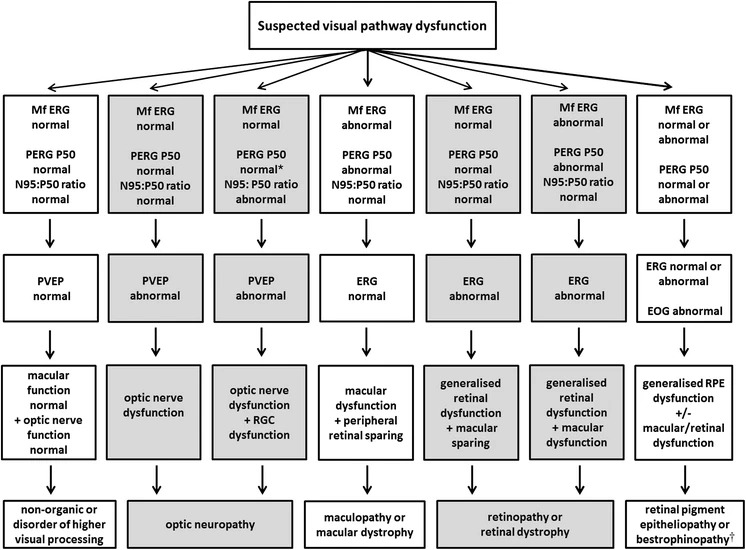
Test Strategy Algorithm¹
Sources
- EyeWiki Electroretinogram
- EyeWiki Electrooculogram
- The Wills Eye Manual: Office and Emergency Room Diagnosis and Treatment of Eye Disease; Kalla Gervasio MD, Travis Peck MD et al; Lippincott Williams&Wilkins; 8th Edition (2021)
- Kanski’s Clinical Ophthalmology: A Systematic Approach; John E Salmon MD; Elsevier; 9th Edition (2019)
- ¹Robson, A.G., Nilsson, J., Li, S. et al. ISCEV guide to visual electrodiagnostic procedures. Doc Ophthalmol 136, 1–26 (2018). https://doi.org/10.1007/s10633-017-9621-y
- International Society for Clinical Electrophysiology of Vision – iscev.org
- American Academy of Ophthalmology; 2017-2018 Basic and Clinical Science Course (BCSC), Section 12 Retina and Vitreous