Change Language German
Medical History
- When was the lesion first noticed?
- Has it changed (size, pigmentation)?
- Personal and family history (sun exposure, skin cancer)?
Examination
- Thorough slit lamp examination of both eyes, including lid eversion
- Localisation, size, thickness, demarcation
- Photo documentation
- Anterior Segment-OCT, ultrasound (depth, scleral involvement?)
Conjunctival Naevus
- Often present since childhood, pigmentation may change during puberty or pregnancy
- Usually unilateral
- Well-demarcated, often with epithelial cysts
- Malignant transformation is rare
- Initial photo , follow-up every 6-12 months
- Consider excision if size, pigmentation, or vessels are suspicious
Complexion-associated Melanosis (CAM)
- Primarily in dark-skinned individuals, may increase with age
- Mostly bilateral, flat, diffuse, and not clearly demarcated, often near the limbus
- Very rarely undergoes malignant transformation
- Initial photo, follow-up every 6-12 months
Congenital ocular Melanocytosis
- Ota naevus when involving the skin (also called Melanosis oculi or Oculodermal melanocytosis)
- Congenital unilateral flat grayish lesion of the sclera and uvea, about 2mm from the limbus, often with periocular bluish skin pigmentation, usually no pigment in the conjunctiva
- More common in African and Asian populations
- Risk of uveal (not conjunctival!) melanoma is about 1:400
- Increased risk of glaucoma
- Follow-up every 1-2 years for melanoma and glaucoma screening
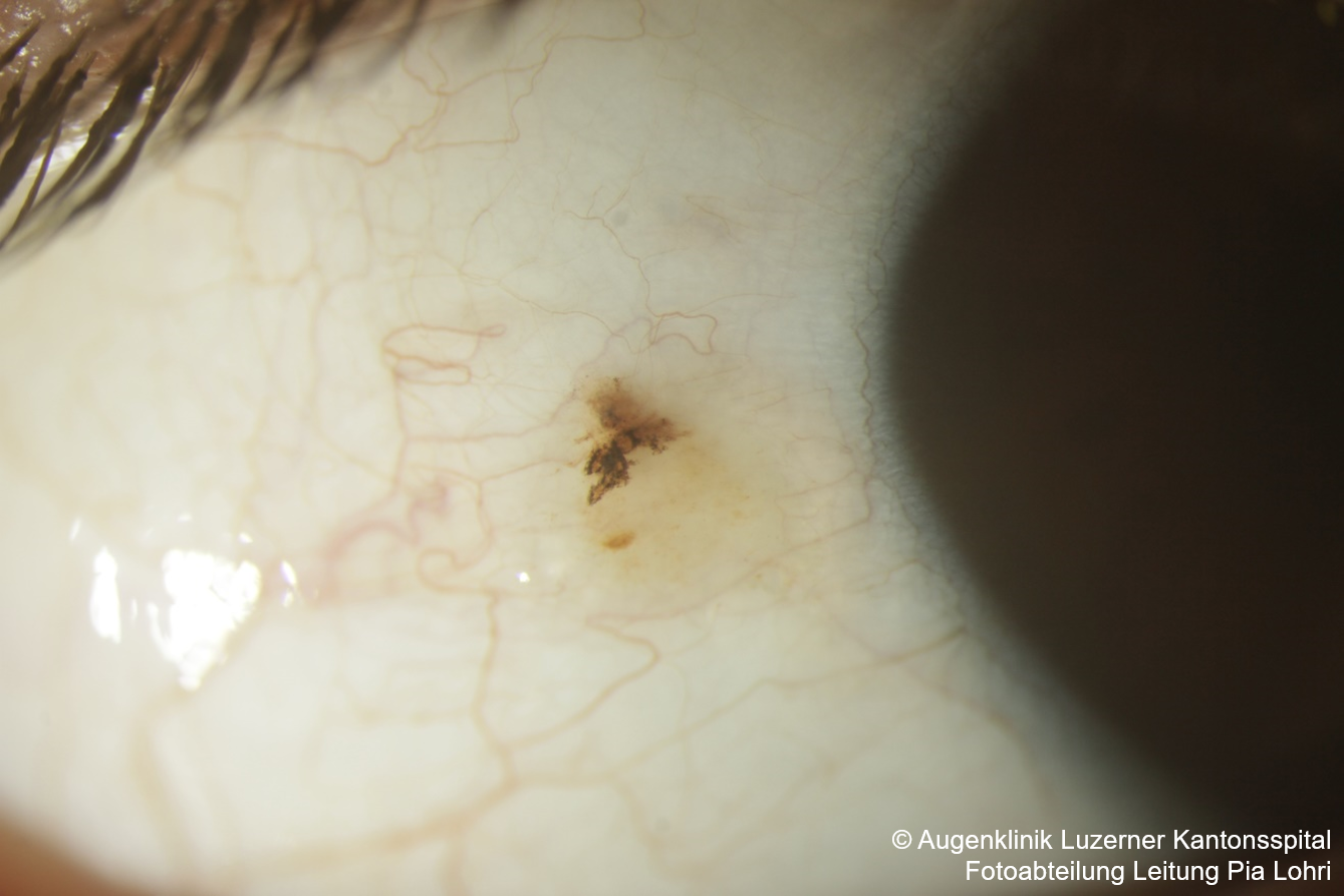
Primary Acquired Melanosis (PAM)
- Newly acquired pigmentation in fair-skinned individuals of middle age (or older)
- If present since childhood, it is more likely a naevus
- Unilateral, flat, speckled golden/yellowish to brown pigmentation, poorly demarcated, non-cystic
- Can also affect the cornea
- Can occur with or without atypia
- With atypia, up to 50% risk of developing melanoma!
- Without atypia, the risk is very small
- Differential diagnosis: naevus
- cysts, well-demarcated, often thicker than PAM
- Treatment
- For small lesions (1-2 clock hours), regular monitoring (6-12 monthly) as long as stable
- If nodules, thickening, or changed vessels, complete excision is indicated!
- For medium-sized lesions (2-5 clock hours), complete excision + cryotherapy of the edges
- For large lesions (>5-6 clock hours), excise thickened or suspicious areas + “map biopsy” of all quadrants
- Consider postoperative topical Mitomycin C (0.02% or 0.04%) if the lesion cannot be completely removed
- For small lesions (1-2 clock hours), regular monitoring (6-12 monthly) as long as stable
Conjunctival Melanoma
- usually arises from PAM with atypia or de novo, transformation from naevus possible
- Most commonly at the limbus, but also at the caruncle, tarsus, fornix
- Non-limbal melanomas have a worse prognosis!
- de novo has the worst prognosis
- Elevated mass, often with feeder vessels, typically brownish, can also be amelanotic
- Metastasis workup
- Excision with no-touch technique
- No incision biopsy!
- Consider sentinel lymph node biopsy for lesions >2mm or high risk
- Prognosis
- Depends on the type and location
- Local recurrences are common (up to 45% at 5 years, 59% at 10 years)
- Mortality at 5-17% at 5 years and 9-35% at 10 years
- ca. 1/3 with metastases after 15 years
Differential diagnoses to PAM or melanomas
- Pingueculum, pterygium, inflammatory granuloma, amyloidosis, Axenfeld loops
- Foreign bodies (e.g., mascara in the inferior fornix, gunpowder after explosions), Silver deposits (Argyrol eye drops), Adrenochrome pigment in the inferior fornix due to epinephrine eye drops
- Ochronosis pigmentation at muscle insertions and in pingueculum in alkaptonuria patients
- Hemorrhagic conjunctival cysts after surgery
- Pigment cells in non-melanocytic tumors
- Calcified Cogan sclera plaque at horizontal rectus muscle insertions in older patients
Sources
- AAO Ophthalmic Pearls Oncology – Conjunctival Pigmented Lesions: Diagnosis and Management
- EyeWiki – Conjunctival Melanocytic Tumors
- EyeWiki – Oculodermal Melanocytosis (Nevus of Ota)
- Shields CL, Shields JA. Tumors of the conjunctiva and cornea. Indian J Ophthalmol 2019;67:1930-48
- Kanski’s Clinical Ophthalmology: A Systematic Approach; John E Salmon MD; Elsevier; 9th Edition (2019)
- Review of Ophthalmology: Neil J. Friedman; Peter K. Kaiser; William B. Trattler; Elsevier, 3rd Edition (2018)